
Reproductive Organ Examination
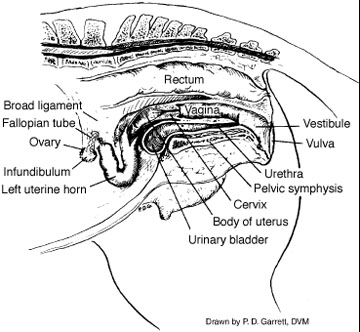
Palpate the mammary system for evidence of mastitis, abscessation, neoplasia or injury. Before examining the external reproductive organs, wash and disinfect the mare’s perineal area and wrap the tail. Examine the vulva for conformation, apposition, tone and evidence of discharge. Poor conformation of the vulva lips and vulva itself will predispose the mare to problems like pneumovagina (windsucking) and fecal contamination of the vagina. Gently separate the vulva lips and listen for the passage of air into the vagina. If you hear air, that is an indication of pneumovagina. The examination should continue onto the clitoris and clitoral fossa. The clitoris has 3 sinuses that have been shown to house the contagious equine metritis (CEM) organism. If CEM is suspected, culture these sinuses. The ovaries, kidney bean in shape, are suspended in the abdomen by a part of the board ligament. Upon palpation, the ovaries must be distinguished between fecal balls in the small colon. The size of the ovaries and the structures present will vary depending on the season. A normal ovary should fit in the palm of your hand and can be examined by the tip of the thumb and fingers. Normal follicles growing on the ovary will range between 2 to 6 cm and should not be confused with cystic structures. Ovulation in the mare occurs at an indentation in the ovary called the ovulation fossa. Examine the pelvis for any structures that might interfere with breeding or parturition. The cervix is the passageway from the vagina to the uterus and can be rectally palpated across the floor of the pelvis. The cervix is a cylindrical structure and is about 5 to 8 cm long.
Mare Reproductive organs
Rectal Examination
With a rectal exam, we can determine if the mare is pregnant before performing the vaginal exam. The rectum of the horse is easily perforated so the veterinarian must be cautious when palpating. The mare’s uterus is T-shaped with 2 short uterine horns and a body. The uterine horns extend upward toward the ovaries. The uterine body is palpated via the rectum approximately 45 to 50 cm into the rectum. The uterus will curve up toward the ovaries, thus allowing you to differentiate it from the small colon. Palpate the uterus for pregnancy or any abnormalities that might be present. Then examine the uterine horns for soundness.
Vaginal Examination
After the perineal area has been thoroughly washed, disinfected, rinsed off and the tail wrapped, a vaginal speculum can be introduced into the vagina. Once fully inserted, the speculum, with the aid of a light, can be used to visually examine the vagina and os of the cervix. The vaginal walls should be examined for color, evidence of inflammation, tumors, lacerations and/or scars. The cervical os should be examined for color and tone as soon as the speculum is fully inserted since changes will occur as cool air enters through the speculum. Remember, the stage, color and shape of the cervix will depend on the stage of the mare’s estrous cycle. Some veterinarians will obtain endometrial uterine cultures through the use of a vagina speculum. An endometrial biopsy can reveal problems within the uterus that might not be detected by palpation. Hormonal analysis for progesterone, estrogen and/or testosterone may be necessary to differentiate causes of enlarged ovaries. In determining the breeding soundness of a mare, all of these examinations may not be required. The type and number of examinations will depend on the reason for the examination, stage of cycle, status of reproductive tract and individual examiner. The abnormalities found should be categorized according to reproductive organs involved or those with evidence of potential abnormalities. The comprehensiveness of a breeding soundness exam will most likely be influenced by time, economics and availability of a qualified examiner.
Kathy Anderson, Extension Horse Specialist, University of Nebraska