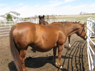
A complete breeding soundness examination should be conducted before you purchase a mare for breeding or the breeding season. This exam should include more than just an evaluation of the reproductive tract, but also a complete history and physical examination of the mare.
History
In evaluating a mare for breeding, it is important to know her general medical and management history plus a detailed reproductive history. A general history would include:
- age
- purchase date
- performance history
- serologic tests
- vaccination history
- boarding facilities
- feed
- previous use
- intended use
- medical history
- surgical history
- disease problems
- weight loss or gain
The reproductive history should include:
- age at first heat
- heat dates
- interval between heats
- length of heats
- age first bred
- breeding dates
- foaling dates
- date of last foaling
- abnormal or assisted foalings
- number of pregnancies
- abnormal pregnancy
- previous year’s breeding cycle pattern
- number of breedings for conception
- evidence of vaginal discharge
- mothering ability
- milk production
- teasing method
- breeding method (pasture, hand breeding or artificial insemination)
An in-depth history review often reveals poor breeding management that results in a mare’s apparent infertility. This situation is easily corrected and is usually a problem with novice breeders. Even though it may appear to be a management problem, a complete physical and reproductive examination should be performed to substantiate your conclusion or isolate other problems.
Physical Examination
Physical examinations are often overlooked in a breeding soundness examination. Basic conformation should be considered to determine if the mare is sound enough to withstand the act of breeding and that she will not pass on any severe conformational abnormalities. Hoof and leg abnormalities, like acute laminitis or joint disorders (osteoarthritis, bursitis), may make her reluctant to stand for breeding. Pelvic injuries or abnormalities may also pose foaling problems. Other conditions may also contribute to breeding and foaling problems. Teeth should also be examined to detect over- and undershot jaws. You should strongly consider not breeding mares with these conditions.
Reproductive Examination
Since mares are seasonal polyestrous and are erratic in their cycles during the transition period from anestrus to the breeding season, we must evaluate the reproductive tract and associated structures in relation to the time or stage of the breeding season.
During anestrus, the uterus is flaccid, thin walled and quiescent. The ovaries are small and firm and the vagina is pale and dry. The cervix is usually in the top portion of the vaginal cavity and is pale, dry and tightly closed. The reproductive tract and findings on the ovaries may be contradicting during the transition period and difficult to predict. However, as the mare begins to cycle, the reproductive organs undergo dramatic changes. As estrogen begins to be the dominant hormone produced by the follicle, the uterus becomes thicker, more vascular and heavier. The cervix begins to move down from the upper portion of the vaginal cavity and starts to have a pinkish color, also getting softer and moister (Figure 1). The cervix at this time may allow the insertion of one finger.

Figure 1: Cervical Changes during estrous cycle.
Figure 1: (1) Diestrus cervix 10 days after ovulation. (2) Onset of estrus. The cervix is swollen and beginning to open. The folds are less defined. (3) Towards the end of estrus, the cervix is swollen and relaxed. The fold hangs down, appearing very vascular. (4) Pregnant cervix, very hard, tightly closed and covered with pasty mucus.
As the estrogen level continues to rise, the cervix continues to drop below the vaginal cavity’s midway point and becomes very soft and pink. Two to three fingers will easily pass through the cervix at this time. Near the time of ovulation and as estrogen peaks, the cervix will be at its lowest point in the vaginal cavity. It may actually be “pooling” on the vaginal floor and be almost indistinguishable. The cervix at this time will have numerous folds and be very soft, reddish pink and extremely moist, while the vaginal walls will be purplish-red, very vascular and moist.
After ovulation, the cervix will return to its original position at the top of the vaginal cavity and will be pale, dry and tightly closed. The “capped” cervix indicates pregnancy (Figure 1). That is, the cervix appears to be covered by one of its folds and its external os cannot be seen.
Reproductive Organ Examination

Figure 2: Reproductive Anatomy of the Mare
Palpate the mammary system for evidence of mastitis, abscessation, neoplasia or injury. Before examining the external reproductive organs, wash and disinfect the mare’s perineal area and wrap the tail. Examine the vulva for conformation, apposition, tone and evidence of discharge (Figure 2). Poor conformation of the vulvar lips and vulva itself will predispose the mare to problems like pneumovagina (windsucking) and fecal contamination of the vagina. Gently separate the vulvar lips and listen for the passage of air into the vagina. If you hear air, that is an indication of pneumovagina.
The examination should continue onto the clitoris and clitoral fossa. The clitoris has 3 sinuses that have been shown to house the contagious equine metritis (CEM) organism. If CEM is suspected, culture these sinuses.
The ovaries (Figure 2), kidneybean in shape, are suspended in the abdomen by a part of the board ligament. Upon palpation, the ovaries must be distinguished between fecal balls in the small colon. The size of the ovaries and the structures present will vary depending on the season. A normal ovary should fit in the palm of your hand and can be examined by the tip of the thumb and fingers. Normal follicles growing on the ovary will range between 2 to 6 cm and should not be confused with cystic structures.
Ovulation in the mare occurs at an indentation in the ovary called the ovulation fossa. Examine the pelvis for any structures that might interfere with breeding or parturition.
The cervix (Figure 2) is the passageway from the vagina to the uterus and can be rectally palpated across the floor of the pelvis. The cervix is a cylindrical structure and is about 5 to 8 cm long.
Rectal Examination
With a rectal exam, we can determine if the mare is pregnant before performing the vaginal exam.
With a rectal exam, we can determine if the mare is pregnant before performing the vaginal exam. The rectum of the horse is easily perforated so the veterinarian must be cautious when palpating.
The mare’s uterus is T-shaped with 2 short uterine horns and a body (Figure 2). The uterine horns extend upward toward the ovaries. The uterine body is palpated via the rectum approximately 45 to 50 cm into the rectum. The uterus will curve up toward the ovaries, thus allowing you to differentiate it from the small colon. Palpate the uterus for pregnancy or any abnormalities that might be present. Then examine the uterine horns for soundness.
Vaginal Examination
After the perineal area has been thoroughly washed, disinfected, rinsed off and the tail wrapped, a vaginal speculum can be introduced into the vagina. Once fully inserted, the speculum, with the aid of a light, can be used to visually examine the vagina and os of the cervix. The vaginal walls should be examined for color, evidence of inflammation, tumors, lacerations and/or scars. The cervical os should be examined for color and tone as soon as the speculum is fully inserted since changes will occur as cool air enters through the speculum. Remember, the stage, color and shape of the cervix will depend on the stage of the mare’s estrous cycle. Some veterinarians will obtain endometrial uterine cultures through the use of a vagina speculum. An endometrial biopsy can reveal problems within the uterus that might not be detected by palpation. Hormonal analysis for progesterone, estrogen and/or testosterone may be necessary to differentiate causes of enlarged ovaries.
Perineal inspection
Poor perineal conformation predispose to “air sucking”, fecal bacterial contamination, and pooling of urine in the vagina. In order to avoid this, the position of the vulva should be close to vertical and approximately ¾ of its length should be below the floor of the pelvis. The vagina is further protected from contamination by a circular sphincter (vestibule-vaginal fold) just inside the urethra. The competency of this sphincter can be assessed by manually parting the lips of the vulva. The sound of aspiration of air into the vagina suggests that the sphincter is compromised, and the mare may need surgical correction for this problem. Other findings of importance to breeding soundness are lacerations, scarring, tumors, and herpes virus lesions on the lips of the vulva.
Uterine culture and cytology
Uterine culture and cytology is obtained to determine if the mare is suffering from a uterine infection. It is important to obtain, process, and evaluate the sample correctly in order for the culture to provide valuable information about the mare’s reproductive health. If a uterine culture is obtained by the use of a swab sample, it should be taken when the mare is in heat. False-negative culture results may occur when samples are obtained in mares out of heat. Uterine cultures from mares that are out of heat can be obtained from biopsy tissue samples. The danger of misdiagnosing an infection based on the growth of a sample contaminant should be recognized, and the culture results should always be performed and interpreted in conjunction with a cytology sample. The cytology sample is obtained from the same swab, or a second swab sample. Bacterial growth in the presence of inflammatory cells is strongly suggesting a uterine infection. In contrast, bacterial growth without any inflammatory cells does most likely reflect contamination of the sample. The presence of inflammatory cells without bacterial growth indicates a non-infectious cause of inflammation such as urine pooling.
Uterine biopsy
A microscopic examination of a tissue sample from the uterine lining provides a valuable piece of information in evaluating the condition of the uterus. The biopsy sample can be obtained at any stage of the cycle and is very harmless to the mare. A biopsy instrument is inserted through the cervix into the uterus and a small piece of the endometrial lining is collected. The biopsy needs to be prepared and stained in a laboratory before it can be evaluated. Interpretation of the biopsy is based on inflammatory and degenerative changes. The presence of inflammatory cells, the health and activity of uterine glands, and the health of the lymphatics are all evaluated and form the basis for a scoring system into four biopsy categories (1, 2a, 2b, and 3). The uterine biopsy grading system can be translated to the mare’s chance to conceive and carry a foal to term.
Summary
In determining the breeding soundness of a mare, all of these examinations may not be required. The type and number of examinations will depend on the reason for the examination, stage of cycle, status of reproductive tract and individual examiner. The abnormalities found should be categorized according to reproductive organs involved or those with evidence of potential abnormalities. The comprehensiveness of a breeding soundness exam will most likely be influenced by time, economics and availability of a qualified examiner.
Resources
Troedsson, M.H.T., Breeding Soundness Examination of the Mare. 1-3.
The Mare: Breeding Soundness Examination and Reproductive Anatomy. 1-4.


